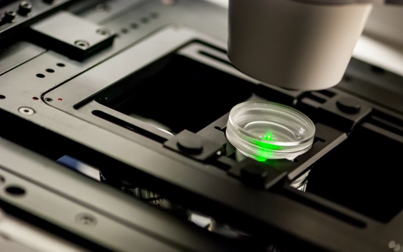
Background: confocal microscopy
Confocal microscopy is a method where illumination is limited to small, diffraction limited spots, resulting in improved resolution. In a classical, single spot confocal microscope, a bright spot is focused on the object, with the scattered light from the object going through a pinhole, to block out-off focused scattered light. This results in a very sharp image of the spot on the object, that by scanning and integration results in a full field image with enhanced resolution compared to a standard light microscope.
Unfortunately, this process of scanning the spot means there is inherent trade off between the imaging speed, the field size and the resolution. To achieve faster imaging, clever illumination schemes are used, often utilizing micro-optical components such a diffractive optical elements and micro lens arrays. A method that is gaining popularity with the increasing availability of laser power is to use a diffractive laser beam splitter to generate an array of diffraction limited spots on the entire view field of the objective. This array can then be imaged in a manner similar to single dot scanning confocal microscopy, resulting in high resolution over the entire field, with short acquisition times.

What is a diffractive laser beam splitter?
A diffractive laser beam splitter is a flat, thin optical component that generates an array of beams from an input laser beam. The beams in the array have defined, equal separation angles, and when focused by an objective create a matrix of dots with pre-defined separations ands arrangement.
For each split laser beam in the matrix, the spot size of the laser beam in the work plane is the same as the diffraction limited spot size of the original laser beam. Thus, there is no reduction in resolution when using a diffractive beam splitter, unlike some cases with lens arrays where aperture diffraction of the sub-lenses can increase the spot diameter of the laser beam at the focal plane.
Laser beam splitter typical use cases in microscopy
Typically, confocal microscopy utilizes wet- immersion objectives with magnification of X60 – X100. Typical laser beam diameters on the objective are ¬6mm, with a resulting diffraction limited spot size of less than 1µm.
View fields are often larger than a hundred microns, requiring splitting angles of 50mrad or more. These are easy achievable with Holo/Or’s diffractive laser beam splitters.
A typical large field microscopy setup consists of a laser beam incident on a laser beam splitter, followed by deflection using a galvo and objective, or x-y motion of the objective to scan the field. This enables scanning multiple spots over the entire objective field, reducing the time necessary to achieve the image by a factor equal to the number of spots. To achieve good performance and high SNR over the entire dot matrix, relatively high-power illumination of >100mw needs to be used. Therefore, the diffractive optical laser beam splitter must be made of monolithic, high durability material such as fused silica. This is especially true for cases such as UV based confocal microscopy, where polymers and plastic optics are mostly useless.
Another configuration that utilizes a diffractive laser beam splitter is an upgrade Nipkow-disc like setup. In this setup, a light pattern similar to that generated by a Nipkow disk is produced by a single diffractive optical laser beam splitter. This element can be rotated much more easily compared to a bulky Nipkow disk, while still reaching diffraction limited spot diameter of the laser beam. By rotating the element, the pattern projected through the objective rotates, resulting in automatic axial scanning of the point in the field.
TL; DR - Q&A
How does confocal microscopy work?
In confocal microscopy, a small, diffraction limited laser dot illuminates a small area in the image field, and the scattered light is filtered to remove high angles. This small spot is scanned over the image field, and results in sharper images compared to illuminating the entire field.
What is a diffractive laser beam splitter?
a diffractive laser beam splitter is a thin, flat component that splits the laser beams in into a matrix of sub-beams with equal separations. When focused, this creates an array of dots.
What is the advantage in using a diffractive laser beam splitter in confocal microscopy?
By generating a matrix of diffraction limited spots, a diffractive laser beam splitter enables the system to scan a much shorter distance, as multiple, separate spots are illuminated at the same time. This significantly reduces the time needed to generate a full image.

